doi: 10.62486/agmu202469
CASE REPORT
Clinical experience of a patient with hemopericardium
Experiencia clínica de un paciente con hemopericardio
Diego
Ernesto Suárez López1 ![]() , Angel
Echevarría Cruz2
, Angel
Echevarría Cruz2 ![]() *
*
1Universidad de Ciencias Médicas de Pinar del Río. Hospital General Docente “Abel Santamaría Cuadrado”. Pinar del Río, Cuba.
2Universidad de Ciencias Médicas de Pinar del Río. Facultad de Ciencias Médicas “Dr. Ernesto Che Guevara de la Serna”. Pinar del Río, Cuba.
Cite as: Suárez-López DE, Echevarría-Cruz A. Clinical experience of a patient with hemopericardium. Multidisciplinar (Montevideo). 2024; 2:69. https://doi.org/10.62486/agmu202469
Submitted: 24-11-2023 Revised: 18-03-2024 Accepted: 30-08-2024 Published: 31-08-2024
Editor: Prof. Dr. Javier
Gonzalez-Argote ![]()
Corresponding author: Angel Echevarría Cruz *
ABSTRACT
Introduction: pericardial effusion is a frequent entity in daily practice, which can occur due to a wide range of pathologies, it is the result of an increase in the blood content of the pericardial sac.
Case presentation: a 93-year-old white female patient came due to a fall from her own feet, was taken to the Orthopedics and Traumatology service where an intertrochanteric right hip fracture was diagnosed and admission to the observation room for subsequent surgical treatment was indicated. The patient began to become tachycardial 110/min, hypotensive 80/50 mmHg, she began to resuscitate with volume responding to mini-fluid challenge (4ml/Kg). The hematocrit is repeated and 0,25 is confirmed, his state of shock is attributed as hypovolemic hemorrhagic. Signs of tissue hypoperfusion persist, despite the prescribed measures and the increase in vasopressor requirements. It was decided to perform an echocardiogram where evident signs of Pericardial Effusion. Pericardial window and hemostasis of the bleeding site were performed, achieving survival of this case.
Conclusions: hemopericardium is an entity with a reserved prognosis, which can occur due to a wide range of traumatic and non-traumatic pathologies; Imaging methods are a key diagnostic tool, the transthoracic echocardiogram is the first-line method for its evaluation. The percutaneous therapeutic approach, surgical exploration, as well as pericardiocentesis are therapeutic methods.
Keywords: Hemopericardium; Cardiovascular Disease; Pericardial Effusion; Cardiac Tamponade.
RESUMEN
Introducción: el derrame pericárdico es una entidad frecuente en la práctica diaria, que puede ocurrir por un amplio rango de patologías, es resultado de un incremento en el contenido de sangre del saco pericárdico.
Presentación del caso: paciente femenina, piel blanca, de 93 años, acude por caída de sus propios pies, es llevada al servicio de Ortopedia y Traumatología donde se Diagnóstica fractura de cadera derecha intertrocantérea y se indica ingreso en sala de observación para posterior tratamiento quirúrgico. La paciente se comienza a tornar taquicárdica 110/min, hipotensa 80/50 mmHg, se comienza a reanimar con volumen respondiendo a minireto de fluídos (4ml/Kg). El Hematocrito se repite y se constata 0,25, se atribuye su estado de Shock cómo Hipovolémico hemorrágico. Persiste los signos de Hipoperfusión tisular, a pesar de las medidas pautadas y del aumento de los requerimientos del vasopresor. Se decide realizar ecocardiograma donde se evidenció signos evidentes de Derrame Pericárdico. Se realiza Ventana Pericárdica y Hemostasia del sitio de sangrado, logrando la supervivencia de este caso.
Conclusiones: el hemopericardio es una entidad de pronóstico reservado, que puede ocurrir por un amplio rango de patologías traumáticas y no traumáticas; los métodos por imágenes constituyen una herramienta diagnóstica clave, el ecocardiograma transtorácico es el método de primera línea en para su evaluación. El abordaje percutáneo terapéutico, la exploración quirúrgica, así como la pericardiocentesis constituyen métodos terapéuticos.
Palabras clave: Hemopericardio; Enfermedad Cardiovascular; Derrame Pericárdico; Taponamiento Cardiaco.
INTRODUCTION
Cardiovascular disease is currently the leading cause of death in industrialized countries and is expected to be the leading cause of death in developing countries by 2020. In Cuba, heart disease has been the leading cause of death in the population over 60 for more than four decades.(1)
For many centuries, heart injuries were considered fatal. Today, cardiac trauma remains one of the most lethal injuries. The outcomes of patients with penetrating cardiac injury can range from deadly injuries to arrhythmias that resolve spontaneously. Hemopericardium in trauma is usually due to penetrating cardiac injury. Still, the pericardial sac may fill with blood from large vessels and rupture of the pericardial artery associated with blunt pericardial laceration.(1,2)
In cases with cardiac rupture, death occurs almost instantly due to acute cardiac tamponade and irreversible electromechanical dissociation. In 50 % of cases, the presentation is sudden death; in the remaining percentage, it manifests with signs of cardiac tamponade or cardiogenic shock that responds transiently to hemodynamic support measures.(1,2)
Pericardial effusion (PE) is a common condition in daily practice that can occur due to a wide range of pathologies and can occur in the context of a wide range of clinical conditions, including heart/kidney failure, inflammation/infections, neoplasms, post-trauma, and post-procedures/surgery. PE results from an increase in the content of the pericardial sac, which usually contains between 15 and 50 ml of serous fluid. PE can be classified according to different criteria: composition, time of onset, amount, hemodynamic impact, or distribution. The presence of mild pericardial effusion could be confused with acute pericarditis. Severe pericardial effusion or tamponade is not the usual presentation of acute pericarditis, and the fluid obtained is usually of a serious nature.(1,2)
Imaging methods are a key diagnostic tool in the evaluation of the pericardium. Transthoracic echocardiography (TTE) is the first-line method for evaluating PD, highlighting its ability to assess cardiac function and the patient’s hemodynamic status. Its limitations include operator dependence and a suboptimal acoustic window determined by the patient’s constitutional habit.(3,4)
The presence of cardiac tamponade requires therapeutic percutaneous (drainage) treatment. Due to the high suspicion of erosion, surgical exploration is usually performed in severe but clinically more stable effusions. The finding of hemopericardium during pericardiocentesis necessitates immediate surgical exploration.
García(5) concludes in his study that around 20 % of clinically stable patients with penetrating precordial trauma present with hemopericardium when the pericardial window makes the diagnosis. This proportion is reduced from 30 % to 50 % when the diagnosis is made by ultrasound. A diagnostic approach using minimally invasive techniques is possible. In hemodynamically stable patients with a positive pericardial window, conservative treatment with drainage, irrigation, and confirmation of the absence of bleeding can be carried out within a strict protocol of intraoperative monitoring and close postoperative surveillance.
CASE PRESENTATION
Female patient, white skin, 93 years old, APP with permanent valvular atrial fibrillation for which she is undergoing regular treatment with Digoxin and Warfarin.
This patient came in after falling on her own feet and was taken to the Orthopedics and Traumatology Department. There, she was diagnosed with an intertrochanteric fracture of the right hip and admitted to the observation ward for subsequent surgical treatment. Perioperative tests were performed, with the following results: hematocrit 0,31, leukocytes 7 x 10⁹, platelet count 250 x 109, and INR 6.
Treatment with vitamin K 10 mg IV and fresh plasma at 15 ml/kg was indicated. The patient began to become tachycardic at 110/min and hypotensive at 80/50 mmHg. Resuscitation was started with volume responding to mini-fluid replacement (4 ml/kg). One hour later, tachycardia persisted at 130; BP was 70/40 mmHg, diaphoretic and obnubilate, so admission to the ICU was decided. Where an EKG was performed, revealing atrial fibrillation with rapid ventricular response, without dynamic ST alterations, with a tendency to microvoltage. An abdominal and soft tissue ultrasound revealed no intra-abdominal collection in the retroperitoneal space or the soft tissues.
Hematocrit is repeated and found to be 0,25. Her condition is attributed to hemorrhagic hypovolemic shock and transfusion with two units of packed red blood cells is initiated, but the expected hemodynamic response is not achieved. Therefore, the airway is addressed, and vasoactive support (norepinephrine) is prescribed, initially at 0,2 mcg/kg/min. Signs of tissue hypoperfusion persisted despite the measures taken and an increase in vasopressor requirements to 0,6 mcg/kg/min, so attempts to locate the bleeding site were continued without success.
An echocardiogram was performed, revealing clear signs of pericardial effusion with imminent signs of cardiac tamponade. The patient went into asystolic cardiac arrest, and cardiopulmonary resuscitation was initiated. A subxiphoid puncture was performed (figure 1), and 500 ml of blood was extracted instantly, bringing the patient out of cardiac arrest. He was transferred to the operating room, where blood component transfusion was optimized, as his HTO reached 0,19. Pericardial window and hemostasis of the bleeding site were performed, achieving survival in this case.
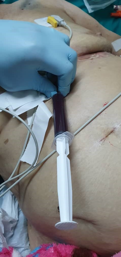
Figure 1. Subxiphoid puncture, instantly extracting 500 ml of blood.
Return to the ICU, where norepinephrine is withdrawn and separation from the VMA is achieved, creating the conditions for hip surgery.
DISCUSSION
In Cuba, heart disease has been the leading cause of death in the population over 60 years of age for more than four decades.(6) Saidman JM et al.(7) state that the presence of blood in the pericardial space can occur in various clinical contexts: iatrogenic, including hemopericardium secondary to invasive cardiac procedures such as percutaneous coronary intervention or pacemaker placement, or as a result of cardiovascular surgery, due to chest trauma caused by high-energy forces leading to the presence of hemopericardium, pericardial rupture, cardiac tamponade, and/or herniation or acute aortic syndrome. In this context, hemopericardium occurs with an estimated frequency of 17-45 %, a fact that contraindicates pericardiocentesis and represents a clinical emergency.
Pericardial pressure depends on the volume of pericardial fluid and its accumulation rate, the phase of the cardiac and respiratory cycle, and the measurement position. The large amount and rapid accumulation of fluid result in a fast and significant increase in pressure.(8)
About traumatic hemopericardium, Morales-Uribe CH et al.(9) state in their study that the diagnostic algorithm in patients with penetrating chest trauma and suspected cardiac injury with hemodynamic stability, recommends performing a FAST (Focused Abdominal Sonography for Trauma), which can achieve a sensitivity of 100 % and a specificity of 96,9 % for detecting hemopericardium. Patients with a positive FAST should undergo sternotomy or thoracotomy for cardiac exploration, as appropriate, while patients with equivocal or inconclusive results should undergo additional diagnostic evaluation, including pericardial window. Patients with a negative FAST and no hemothorax or pneumothorax may be discharged.
Not all cases of traumatic hemopericardium due to stab wounds require thoracotomy. Conservative management with pericardial window, hemopericardial drainage, lavage, and drainage is an option in hemodynamically stable patients who show no evidence of active bleeding after hemopericardial drainage.(9)
Custodio-Marroquín(10) concludes in his study that massive hemopericardium is a rare but potentially fatal complication in cardiac surgery patients. Its prevalence is higher in patients with mechanical prostheses who receive oral anticoagulation. A multidisciplinary approach and systematic follow-up are necessary to maintain the desired anticoagulation range and identify possible complications early.
Echocardiography is the primary diagnostic tool for quantifying the effusion, presence of septa, and functional or hemodynamic repercussions with the collapse of the atrium and right ventricle at the end of diastole.(11)
Drainage by pericardiocentesis was first described in 1955 by Fallows and Pastor. Since then, numerous reports have demonstrated the efficacy and safety of this procedure. Percutaneous pericardiocentesis can be performed blindly in an emergency or guided by transthoracic echocardiography, which allows for safer drainage. This procedure should be performed in the intensive care unit or cardiac catheterization laboratory, where monitoring methods and adequate means of response to possible complications are available, including airway management and cardiopulmonary support.(11)
CONCLUSIONS
Hemopericardium is a condition with a guarded prognosis that can occur due to a wide range of traumatic and non-traumatic pathologies. Imaging methods are a key diagnostic tool, with transthoracic echocardiography being the first-line method for evaluation. Therapeutic percutaneous approaches, surgical exploration, and pericardiocentesis are therapeutic methods.
REFERENCES
1. Piamo-Morales AJ, Ferrer-Marrero D, Palma-Machado L, Arzuaga-Anderson I, Chávez-Jiménez D, García-Rojas Mayra A. Rotura miocárdica de ventrículo izquierdo secundaria a infarto agudo de miocardio. AMC [Internet]. 2019 Jun [citado 2024 Mayo 17] ; 23( 3 ): 349-360. Disponible en: http://scielo.sld.cu/scielo.php?script=sci_arttext&pid=S1025-02552019000300349&lng=es.
2. da Costa Medeiros BJ, Oliveira Araujo A, Daumas Pinheiro Guimarães A. Hemopericardio Por Disparo Sin lesión cardíaca, descripción De Un Mecanismo De Trauma. Rev Colomb Cir. [Internet] 2020 [citado 17/5/2024], 35, 108-112. Disponible en: https://www.revistacirugia.org/index.php/cirugia/article/view/594
3. Saidman JM, Falconi M, Arenaza Pérez D, Gutiérrez Vallecillo MJ, Gentile E, Ulla M. Utilidad de la tomografía computarizada multicorte para la estimación de la composición del derrame pericárdico. Rev. argent. radiol. [Internet]. 2022 [citado 2024 Mayo 17] ; 86( 3 ): 199-210. Disponible en: http://www.scielo.org.ar/scielo.php?script=sci_arttext&pid=S1852-99922022000300199&lng=es
4. Contreras AE, Peirone AR, Ledesma F, Juaneda E, Defago V, Cuestas E. Derrame pericárdico y erosión cardiaca por dispositivos oclusores de defectos interauriculares. Arch. Cardiol. Méx. [Internet]. 2022 [citado 2024 Mayo 17]. 92 (4). Disponible en: https://www.scielo.org.mx/scielo.php?pid=S1405-99402022000400534&script=sci_arttext
5. García A. Enfoque inicial del paciente estable con trauma precordial penetrante: ¿es tiempo de un cambio? Conferencia “Rafael Casas Morales”, XLIV Congreso Nacional “Avances en Cirugía”, Cartagena, Colombia, agosto de 2018. rev. colomb. cir. [Internet]. 2019 Mar [cited 2024 Aug 15] ; 34( 1 ): 16-24. Available from: http://www.scielo.org.co/scielo.php?script=sci_arttext&pid=S2011-75822019000100016&lng=en.
6. Piamo-Morales AJ, Ferrer-Marrero D, Palma-Machado L, Arzuaga-Anderson I, Chávez-Jiménez D, García-Rojas Mayra A. Rotura miocárdica de ventrículo izquierdo secundaria a infarto agudo de miocardio. AMC [Internet]. 2019 Jun [citado 2024 Ago 15] ; 23( 3 ): 349-360. Disponible en: http://scielo.sld.cu/scielo.php?script=sci_arttext&pid=S1025-02552019000300349&lng=es
7. Saidman JM., Falconi M, Arenaza Pérez de D, Gutiérrez-Vallecillo MJ., Gentile E. Utilidad de la tomografía computarizada multicorte para la estimación de la composición del derrame pericárdico. Rev. argent. radiol. [Internet]. 2022 [citado 2024 Ago 15] ; 86( 3 ): 199-210. Disponible en: http://www.scielo.org.ar/scielo.php?script=sci_arttext&pid=S1852-99922022000300199&lng=es.
8. Medeiros Bruno JC, Araujo Oliveira A, Guimarães Daumas A. Hemopericardium by gunshot without cardiac injury, description of a trauma mechanism. rev. colomb. cir. [Internet]. 2020 Mar [cited 2024 Aug 19] ; 35( 1 ): 108-112. Disponible en: http://www.scielo.org.co/scielo.php?script=sci_arttext&pid=S2011-75822020000100108&lng=en.
9. Morales-Uribe CH, López A, Sepúlveda Sandra M. Manejo conservador del hemopericardio por trauma: reporte de un caso. rev. colomb. cir. [Internet]. 2023 Mar [cited 2024 Aug 18] ; 38( 1 ): 195-200. Available from: http://www.scielo.org.co/scielo.php?script=sci_arttext&pid=S2011-75822023000100195&lng=en.
10. Custodio-Marroquín JA. Hemopericardio masivo por sobreanticoagulación en portador de prótesis valvular mecánica: Reporte de caso. Rev. Cuerpo Med. HNAAA [Internet]. 2021 Abr [citado 2024 Ago 18] ; 14( 2 ): 226-228. Disponible en: http://www.scielo.org.pe/scielo.php?script=sci_arttext&pid=S2227-47312021000200018&lng=es.
11. López Masó IM, Ricardo Pérez A. Hemopericardio de oscura etiología en paciente con síndrome de Poland. Cir Ped [Internet]. 2013 [citado 2024 Ago 18] ; Disponible en: http://www.cirugiapediatrica2013.sld.cu/index.php/cirugiapediatricaholguin/2013/paper/viewFile/41/35
CONFLICT OF INTEREST
None.
FUNDING
None.
AUTHORS CONTRIBUTIONS
Conceptualization: Angel Echevarría Cruz.
Data curation: Diego Ernesto Suárez López.
Formal analysis: Diego Ernesto Suárez López.
Fund acquisition: Diego Ernesto Suárez López.
Research: Angel Echevarría Cruz.
Methodology: Angel Echevarría Cruz.
Project management: Diego Ernesto Suárez López.
Resources: Angel Echevarría Cruz.
Software: Angel Echevarría Cruz.
Supervision: Diego Ernesto Suárez López.
Validation: Diego Ernesto Suárez López.
Visualization: Angel Echevarría Cruz.
Writing – original draft: Angel Echevarría Cruz.
Writing – revision and editing: Diego Ernesto Suárez López.